
***Nouvelle date limite: 29 mai 2025***
Centre d’innovation biomédicale, Faculté de médecine, Université de Montréal
Le Centre d’innovation biomédicale (CIB) est une initiative majeure de la Faculté de médecine de l’Université de Montréal. Sa mission est d’explorer les mystères de la biologie humaine afin de décrypter les mécanismes fondamentaux des organismes vivants et de stimuler la découverte de nouveaux traitements.
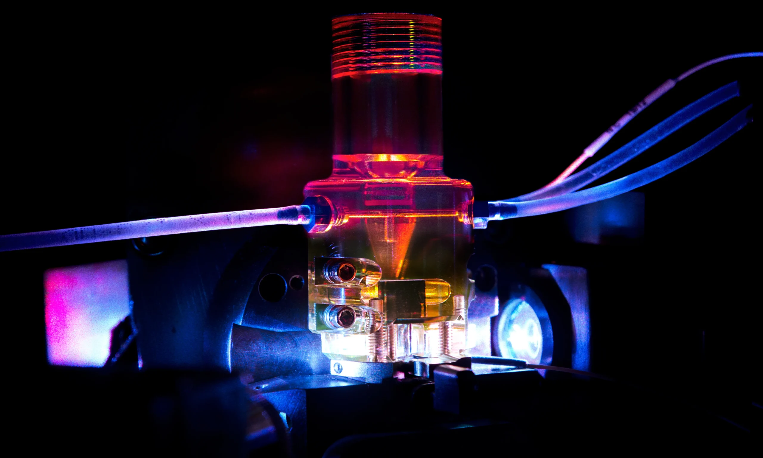
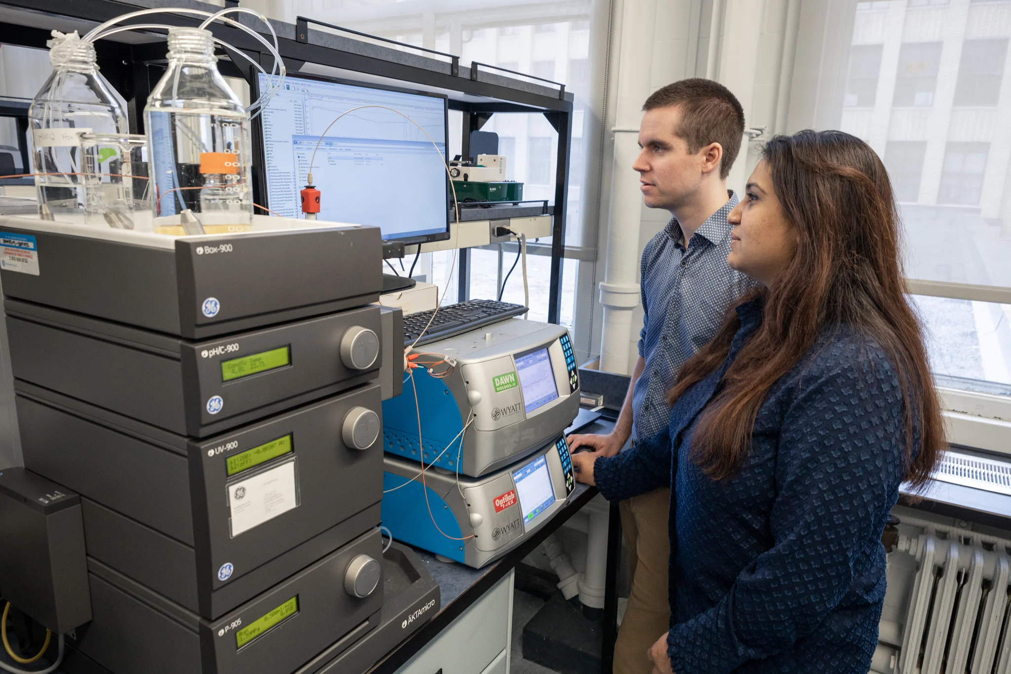
Les 50 équipes du CIB favorisent l’intégration de méthodes quantitatives et d’approches novatrices, en s’appuyant sur des plateformes technologiques de pointe pour mieux comprendre le fonctionnement des organismes vivants, de la molécule unique à leur organisation en systèmes cellulaires complexes. Nos recherches approfondies explorent notamment la physiologie bactérienne et la découverte de nouveaux antibiotiques, la structure des macromolécules et la régulation des gènes et des protéines, la composition cellulaire du cerveau et les neurosciences computationnelles, l’organisation de nos organes et tissus, et bien plus encore. Nous partageons ouvertement nos données, nos idées et nos outils avec la communauté scientifique mondiale afin d’accélérer les progrès et de libérer le plein potentiel de nos découvertes.
Située au Québec, au Canada, Montréal offre un mélange unique de charme européen et de dynamisme nord-américain, avec sa gastronomie de renommée mondiale et une scène culturelle riche, animée par d’innombrables festivals et événements. Au-delà de son atmosphère vibrante, Montréal bénéficie d’un marché du travail florissant, d’un coût de la vie abordable et d’une communauté bilingue et accueillante. De ses parcs et espaces verts magnifiques à sa vie nocturne et sa scène artistique dynamiques, Montréal offre une qualité de vie exceptionnelle.
Le CIB sollicite des candidatures de jeunes stagiaires postdoctoraux talentueux pour un programme de bourse de deux ans dans les domaines étudiés par ses chercheuses et chercheurs.
Objectifs du programme
Ce programme vise à :
- Attirer les meilleurs chercheurs postdoctoraux du monde entier et les futurs leaders dans les domaines et sujets liés aux priorités stratégiques du Centre d’innovation biomédicale (CIB), de la Faculté de médecine de l’Université de Montréal (UdeM).
- Favoriser la rétention des personnes les plus talentueuses issues de nos laboratoires.
- Encourager le développement de projets de recherche collaboratifs et appliqués de pointe.
- Offrir une expérience immersive au sein de l’écosystème de l’UdeM.
Description du financement
Le montant annuel alloué comprend le salaire complet du stagiaire postdoctoral, conformément aux principes établis par l’Université de Montréal. Cela inclut un salaire annuel pouvant atteindre 63 819 $ CA, en plus des avantages sociaux complets.
La durée totale de la bourse est de deux ans maximum.
Cette bourse ne peut être cumulée avec une autre bourse nominative d’un organisme subventionnaire de manière à dépasser le montant maximal de la bourse (par exemple, CRSNG, CRSH, IRSC).
Les candidates et candidats provenant de pays admissibles à l’aide publique au développement du Canada peuvent, sur demande, se faire rembourser leurs frais de réinstallation au Québec, conformément aux règles de ce programme.
Critères d’admissibilité
La candidate ou le candidat doit :
- Avoir obtenu son doctorat (Ph. D.) moins de trois ans avant le début du financement ; ou
- Prévoir obtenir son doctorat avant le début du financement ; et
- Maintenir son statut de stagiaire postdoctoral pendant toute la durée du financement (2 ans).
Les candidatures des groupes sous-représentés sont fortement encouragées*.
La superviseure ou le superviseur doit :
- Être membre régulier de la Faculté de médecine ET du CIB ;
- Occuper l’un des postes admissibles suivants : professeur de recherche, professeur (incluant les professeurs sous octroi et cliniques) au rang d’adjoint, agrégé ou titulaire ;
- Ne pas être ou avoir été le directeur ou le codirecteur de thèse de la candidate ou du candidat.
Processus de candidature
Les candidates et candidats doivent soumettre leur dossier de candidature à postdoc@cib.umontreal.ca.
Les candidatures soumises après la date limite ne seront pas prises en considération.
Le dossier de candidature doit inclure :
- Le formulaire de candidature
- La description de votre projet potentiel (maximum 3 pages ; les références peuvent être listées sur une page supplémentaire) ;
- Une lettre de motivation (maximum 2 pages) expliquant le profil de carrière envisagé et la pertinence de la bourse postdoctorale dans ce contexte.
- Votre CV (format libre, en PDF) ;
- Vos relevés de notes officiels du doctorat (veuillez expliquer le barème de notation dans le cas des universités non canadiennes) ;
- Au moins deux lettres de recommandation ;
- Vous pouvez également inclure une lettre d’appui d’un superviseur admissible, si cette personne a déjà accepté de soutenir votre candidature.
Les candidatures peuvent être soumises en anglais ou en français.
Date limite pour postuler : 29 mai 2025
Des détails supplémentaires, incluant une liste des superviseurs potentiels admissibles et des projets d’intérêt, sont disponibles sur notre site (voir ci-bas).
*Consultez le formulaire de candidature pour les définitions appropriées.
16 Projets disponibles
Les projets décrits plus bas ont été soumis par les chercheuses et chercheurs principaux du CIB. Vous pouvez les contacter afin de discuter de cette opportunité avant de soumettre votre candidature. Vous pouvez également adapter votre candidature en fonction d’un des projets affichés. Chaque projet n’est pas obligatoirement associé à une bourse. L’application sera sujette au même processus d’évaluation selon le financement disponible.
Variantes génétiques de KCNA et KCNB dans les réseaux neuronaux dérivés des iPSC
Superviseur: Rikard Blunck
Les canaux potassiques voltage-dépendants jouent un double rôle dans les neurones : ils repolarisent les membranes après une excitation, et ils régulent l’excitabilité neuronale. En collaboration avec le service de dépistage génétique de l’Hôpital pour enfants Sainte-Justine et des collaborateurs européens, nous étudions les effets des variants génétiques identifiés chez des patients atteints de troubles neurodéveloppementaux et d’encéphalopathie épileptique développementale. L’objectif de ce projet est d’identifier les mécanismes moléculaires sous-jacents à ces variants génétiques par électrophysiologie et fluorométrie sous voltage-clamp, combinées à des simulations de biochimie des protéines et de dynamique moléculaire. Nous utiliserons ensuite des échantillons biologiques de patients pour générer des cellules iPSC ou éditer génétiquement le variant dans des cellules iPSC. L’effet sur la fonction et l’excitabilité des réseaux neuronaux dérivés de cellules iPSC sera étudié par des mesures de patch-clamp double et de réseaux multiélectrodes (MEA).
Ce projet fait partie dans un effort plus vaste visant à identifier des traitements personnalisés pour les maladies rares. À ce titre, le laboratoire fait partie, en plus du Centre d’innovation biomédicale, du Centre de recherche interdisciplinaire sur le cerveau et l’apprentissage (CIRCA) et du Réseau maladies rares Québec (RareQC).
Site web du laboratoire: www.biophys.umontreal.ca/bluncklab
Publication : https://pubmed.ncbi.nlm.nih.gov/38503299/
Une nouvelle génération d’interfaces neurales intelligentes et autonomes pour la neurostimulation
Superviseur: Marco Bonizzato
Ce travail introduit une nouvelle méthode d’optimisation d’interfaces neurales capable d’accélérer de plusieurs ordres de grandeur leur paramétrage et leur déploiement.
Nous avons démontré l’efficacité de l’Optimisation Bayésienne (OB) pour sélectionner les paramètres de neurostimulation dans des modèles animaux (rat, primate non humain), permettant ainsi de maximiser l’output moteur et d’obtenir des améliorations significatives de la locomotion après lésion médullaire.
Toutefois, le coût computationnel de l’OB demeure élevé et augmente considérablement avec le nombre d’itérations. En pratique, la méthode devient rapidement trop coûteuse en ressources pour être viable sur des périodes prolongées ou pour des optimisations extrêmement rapides.
Nous proposons donc d’explorer des méthodes d’apprentissage profond et d’apprentissage par renforcement pour surmonter ces limites. Cette approche soutient le décideur algorithmique OB et lui permet d’identifier efficacement et rapidement les paramètres optimaux de neurostimulation.
Contrairement aux techniques actuelles, longues et coûteuses, cette optimisation accélérée pourrait faciliter considérablement l’application clinique des neuroprothèses.
https://polymtl.ca/expertises/en/bonizzato-marco
Stimulation des circuits de récompense pour restaurer le mouvement après neurotraumatisme
Superviseur: Marco Bonizzato
Ce projet explore, chez le modèle du rat, l’utilisation de la stimulation cérébrale profonde (DBS, Deep Brain Stimulation) ciblant les circuits cérébraux de récompense et d’apprentissage moteur pour favoriser la récupération motrice après neurotraumatisme, notamment les lésions médullaires.
Bien que ces lésions réduisent fortement la mobilité, certaines connexions nerveuses persistent et peuvent être renforcées par réadaptation. Cependant, la récupération reste souvent limitée.
En stimulant spécifiquement les circuits de récompense impliqués dans l’apprentissage moteur, cette approche pourrait immédiatement améliorer la motricité tout en facilitant une récupération durable du contrôle volontaire. Nous utilisons une DBS adaptive et en boucle fermée pour façonner l’exécution du mouvement pendant l’entrainement en réadaptation.
Le projet vise à démontrer l’efficacité de cette modalité de DBS, une technologie utilisée cliniquement pour d’autres indications (Parkinson, dystonie) mais encore inexplorée pour la paralysie. À terme, cette stratégie pourrait devenir un traitement clinique innovant pour les personnes vivant avec une paralysie.
https://polymtl.ca/expertises/en/bonizzato-marco
Exploring the proteome of pathogenic RNAs with nucleotide expansion repeats
Superviseur: Pascal Chartrand
The Chartrand lab is looking for a motivated postdoctoral fellow interested to study the proteome of toxic RNA with nucleotide repeat expansions, like CUG, CAG or CGG, involved in the pathogenesis of several neuromuscular and neurodegenerative diseases, like myotonic dystrophy, amyotrophic lateral sclerosis (ALS) or spinocerebellar ataxias. This project aims to understand how these toxic repeat RNAs can sequester and disrupt the function of proteins involved in RNA metabolism. This project is in collaboration with the lab of Dr. Éric Lécuyer at IRCM (Montréal), and funded by CIHR and Genome Quebec (https://www.ircm.qc.ca/en/news-detail/dr-lecuyer-and-dr-chartrand-winners-of-innovative-therapies-hereditary-ataxias-competition)
The successful candidate will lead pioneering work in the field using high-throughput imaging screen and RNA-based proximity labeling (BioID) assays to identify common RNA-binding proteins associated with nucleotide repeat expansions. Our laboratory also uses leading-edge techniques in single-molecule imaging, RNA and protein biochemistry, cellular models of diseases, and functional genomics to dissect the roles of RNA-binding proteins involved in the pathogenesis of repeat expansion diseases. For more information about the Chartrand lab: https://recherche.umontreal.ca/english/our-researchers/professors-directory/researcher/is/in14535/
Neurophysiological Effects of Repetitive Transcranial Magnetic Stimulation (rTMS) in Preclinical Models of Stroke
Superviseur: Numa Dancause
A postdoctoral position is available in the laboratory of Dr. Numa Dancause at the Université de Montréal,
Department of Neurosciences. Our group investigates the mechanisms underlying movement control, neural
plasticity involved in motor recovery post-brain injury, and the effects of neuromodulation techniques such as
repetitive transcranial magnetic stimulation (rTMS) on the brain. We employ rodent and macaque monkey models, and benefit from collaborations with human-focused research groups through our Canadian platform Can-Stim. Here is a full list of our published work.
The project: The postdoctoral fellow will investigate the effects of rTMS in macaque stroke models, integrating
neural and electromyographic recordings while employing an exoskeleton interface, the KINARM. The project will
involve:
- Conducting neural recordings in behaving monkeys.
- Implementing and analyzing lesion models and neuro-recovery processes.
- Programming in MATLAB or Python for data analysis.
- Collaborating with interdisciplinary neuroscience, engineering, computational and clinical rehabilitation
teams.
Your profile:
- Training in neuroscience, biomedical engineering, behavioral sciences, or a related field.
- Strong programming and electrophysiological data analysis skills, particularly in MATLAB or Python.
- Experience with electrophysiological techniques in vivo AND/OR neuromodulation techniques.
- Experience with behavioral shaping in non-human primates or other animals
- Motivation to pursue innovative research in neurobiology and motor rehabilitation.
- Excellent communication skills and ability to work collaboratively in a research team.
Centre Interdisciplinaire de Recherche sur le Cerveau et l’Apprentissage; CIRCA
Innovation Fellow in Motor Cognition
Superviseurs: Becket Ebitz et Numa Dancause
The Ebitz and Dancause labs in the Department of Neurosciences at the Université de Montréal are recruiting a postdoctoral fellow for a project at the interface of cognitive and motor control. Humans and other animals can produce the same action for multiple reasons. For example, they can grasp because they think it’s the best thing to do, because they’re curious about what will happen, or because they are bored. The goal of this project is to understand how these cognitive states shape the integration of motor control processes in the brain. The project will combine (1) large-scale neural recordings from the motor and premotor cortex with (2) cutting edge computational models to infer the hidden cognitive states underlying action. There are also opportunities for causal perturbation studies that would combine large-scale neural recordings with electrical or transcranial magnetic stimulation. The ideal candidate will be interested in building on the insights from this project to develop technologies for motor rehabilitation.
https://recherche.umontreal.ca/english/our-researchers/professors-directory/researcher/is/in14962/
https://www.youtube.com/watch?v=6np7KNyOe-w&ab_channel=MAINConference
Targeting a novel NAD dependent metabolic complex to treat cancer
Superviseur: Gerardo Ferbeyre
Collaborateurs: Ivan Topisirovic et Malik Chaker-Margot
Cancer treatment is challenging due to similarities between cancer and normal cells. Cancer cells can adapt to stress, including chemotherapy, through metabolic changes. While mutations in cancer genes are traditionally seen as driving carcinogenesis, emerging evidence indicates epigenetic changes in cancer cells, unrelated to these mutations. Our team discovered a metabolic cycle in cells under oncogenic stress, catalyzed by a complex found in advanced prostate and breast cancers. Targeting this complex could offer a new treatment approach. We have already shown that targeting the individual enzymes in this complex using genetic tools show strong anti-neoplastic and anti-metastatic properties in pre-clinical breast cancer models. We aim now to design compounds that disrupt the complex without inhibiting its individual enzymes, based on understanding its biochemical and biophysical principles.
Key References:
Contact: g.ferbeyre@umontreal.ca
Understanding embryonic development in latent space
Superviseur: Paul François
In living systems, all processes are intrinsically dynamic. But, even the most basic biological dynamics are of such high-dimensional character that it is often difficult to deduce representations containing the most essential features with high predictive power. The project’s goal is to develop approaches to build interpretable models of embryonic development, such as vertebrate segmentation or early cell-fate decisions, using tools from dynamical systems theory, theoretical biophysics, and machine learning. Candidates should have a Ph.D. in theoretical or computational physics/biology/biophysics/computer science, or a related field, and a strong interest in biology. Experience in machine learning techniques (in a broad sense) is a plus. The candidate will be part of a highly interdisciplinary team of theoretical biophysicists and bioinformaticians, and the project will be done in close collaboration with experimentalists.
Relevant links :
Francois Research Group : https://www.francoisresearch.org/
Introductory article on latent space modelling developed in our group : https://www.science.org/doi/10.1126/science.abq1679
Review on vertebrate segmentation : https://arxiv.org/abs/2403.00457
The role of astrocytes in axonal information processing
Superviseure: Arlette Kolta
Neurons can be conceptualized as input-output systems, receiving, integrating, and processing information before generating an output signal. Neuronal communication depends on the reliable and efficient propagation of action potentials (APs) along the axon. Long regarded as a simple electrical cable whose sole function was to transmit AP trains; the axon is now recognized as a dynamic and complex structure. It exhibits morphological plasticity and is sensitive to both internal and external factors that can modulate AP waveforms (amplitude, width) and thus alter the amount of information transmitted. Among these factors, astrocytes have emerged as potential modulators of axonal transmission, notably through the release of gliotransmitters. S100β, a calcium-binding protein, is of particular interest due to its role in regulating extracellular calcium homeostasis and several voltage-gated channels expressed at all levels in the axonal compartment and involved in AP waveform modulation. This project seeks to understand how the gliotransmitters S100β modulate axonal transmission in the layer 5 pyramidal neurons (L5PN) of the visual cortex using a combination of high-end in vitro electrophysiology (bleb recording, paired recording), calcium and voltage imaging and immunohistochemistry.
Financement: https://webapps.cihr-irsc.gc.ca/decisions/p/project_details.html?applId=509122&lang=en
https://www.frontiersin.org/journals/cellular-neuroscience/articles/10.3389/fncel.2024.1477985/full
https://recherche.umontreal.ca/english/our-researchers/professors-directory/researcher/is/in13874/
Role of brainstem circuits in producing complex hand to mouth movements
Superviseure: Arlette Kolta
Feeding is a complex behavior which execution and modulation depend on integration of sensory, motor and limbic inputs by brainstem and spinal circuits coordinating jaw, tongue, facial and limb muscles. Mastication, which represents the rhythmic jaw movements responsible for breaking down ingested food to prepare it for digestion, is central to feeding. As for other rhythmic movements, this process is thought to be controlled by a neuronal network, called Central Pattern Generator, located primarily in the brainstem in this case.
However, feeding is more than just mastication and relies in many species on hand-to-mouth coordination. The sensori-motor integration required for this coordination is thought to occur in the motor cortex and to involve higher centers such as the cerebellum. However, recent unpublished data from our laboratory suggest that focal optogenetic stimulation of some trigeminal brainstem nuclei that are part of the masticatory CPG, can produce coordinated hand-to-mouth movements raising the possibility that complex motor behavior may be entirely represented at the level of the brainstem and the spinal cord. This project explores this hypothesis using optogenetics and viral tract tracings techniques in transgenic mice lines.
Financement: https://webapps.cihr-irsc.gc.ca/decisions/p/project_details.html?applId=322941&lang=en
https://recherche.umontreal.ca/english/our-researchers/professors-directory/researcher/is/in13874/
Contribution of astrocytes to changes in large primary afferents excitability associated to chronic muscle pain
Superviseure: Arlette Kolta
Muscle pain is associated to trigger points in many syndromes like fibromyalgia and with small spots when induced, with needles for EMG studies for instance. These spots are often associated to spontaneous electrical activity that seems to emanate from fibres inside muscle spindles. These observations, added to the reports that large diameter primary afferents (PAs) like those innervating muscle spindles become hyperexcitable and develop spontaneous ectopic firing in conditions leading to neuropathic pain, suggest that excitability changes of these afferents may contribute importantly to development of pathological pain. This project explores the cellular and ionic mechanisms leading to the development of this hyperexcitability and to ectopic firing with a particular emphasis on the contribution of neuron-glia interaction. Our previous work has shown the impact of an astrocytic Ca2+-binding protein, S100β, on neuronal functions supported by a persistent sodium current (INaP) implicating Na+ channels containing the a subunit Nav1.6. These channels play a crucial role in generating firing and regulating excitability of large diameter PAs. This project investigates the hypothesis that large diameter PAs hyperexcitability results from increased astrocytic (or satellite cells) activity leading to an increased S100β-mediated enhancement of function of Nav1.6 channels, using immunohistochemistry, electrophysiology, and Ca2+-imaging techniques, combined to optogenetic manipulations of astrocytes in transgenic mice lines.
Financement: https://webapps.cihr-irsc.gc.ca/decisions/p/project_details.html?applId=402085&lang=en
https://recherche.umontreal.ca/english/our-researchers/professors-directory/researcher/is/in13874/
Systematic design of cis-regulatory elements along developmental transitions
Superviseur: Jean-Benoît Lalanne
Regulatory elements are DNA fragments of the genome that respond to the biochemical state of cells to control expression of genes in space and time within complex organisms. The resulting specificity of gene expression control (where, when, how much) is thus encoded in the DNA sequence. Most previous quantitative experimental and computational work has focused on understanding differences in regulatory element function either within a fixed cellular state (e.g., in one cell line), or across highly distinct, discrete cellular states (e.g., different cell-types or lines). However, along differentiation trajectories, cells can occupy a continuum of intermediate states. Importantly diseased cells, such as some cancer cells, often only partially complete developmental transitions and display such hybrid character. This provides a strong biomedical rationale to design regulatory elements that can precisely detect such intermediate states. Leveraging genomics method development, high-content single-cell functional profiling, predictive mathematical modeling, and synthetic biology, this project aims to engineer regulatory element ‘sensors’ that respond uniquely to hybrid cellular states along developmental trajectories. Accomplishing this wedge goal requires pushing the limit of our quantitative understanding of the gene-regulatory code, and will leverage the lab’s newly developed single-cell-resolution barcoded enhancer method. The ideal candidate would have strong expertise in both experimental and computational genomics, mammalian cell culture, and molecular biology.
Links:
Lab website: lalannelab.org
Summary of our new method: https://www.nature.com/articles/s41592-024-02261-2
Structural Characterization of Regulatory Mechanisms Controlling let-7 microRNAs in Development and Cancer
Superviseur: Pascale Legault
MicroRNAs (miRNAs) are small, non-coding RNAs that post-transcriptionally regulate gene expression by binding to the 3′ untranslated region (3’UTR) of target mRNAs, promoting their degradation or translational repression. The let-7 family is a well-characterized and conserved group of miRNAs with critical roles in development, cell differentiation, and tumor suppression. Despite their biological importance, the mechanisms underlying the maturation and regulation of let-7 miRNAs remain poorly understood.
Our laboratory has recently identified a set of candidate proteins that bind to the stem-loop structures of immature let-7 precursors, using affinity purification and mass spectrometry. These proteins form a densely interconnected protein–protein interaction network, suggesting the presence of macromolecular assemblies involved in the post-transcriptional regulation of let-7 biogenesis.
This postdoctoral project aims to elucidate the structural basis of protein-mediated regulation of let-7 miRNAs. Specifically, we will (1) reconstitute RNA–protein complexes in vitro, (2) characterize their composition and dynamics using biochemical and biophysical approaches (EMSA, enzymatic assays, mass photometry, ITC, etc.) and (3) determine their structure via cryo-electron microscopy (cryo-EM). By integrating structural data with functional assays, this work will uncover how specific proteins and protein complexes influence let-7 maturation and activity.
The successful candidate will join a dynamic and collaborative research team with strong expertise in RNA biology and structural biochemistry within the new Center for Biomedical Innovation, providing an excellent environment to support and advance their work. These studies will provide mechanistic insights into miRNA regulation and may reveal novel therapeutic targets relevant to development and cancer.
https://recherche.umontreal.ca/chercheur/is/in14566/
http://airen.bcm.umontreal.ca/
The Paradox of Parasitism: Evolutionary Dynamics of Nested Parasites in Marine Systems
Superviseur: Frédérique Le Roux
How can parasitism—a relationship where one organism harms or kills its host—persist in nature? Several mechanisms offer explanations: opportunistic or generalist pathogens, antagonistic coevolution with trade-offs, and even hyperparasitism, where parasites themselves are parasitized. In our lab, we explore this paradox through a marine model involving invertebrate pathogens (vibrios), their phages, and phage satellites. We host extensive collections of natural Vibrio populations and their phages, exhibiting striking genome size variation linked to lifestyle differences. As a postdoctoral fellow in bioinformatics, you will work with >600 Vibrio genomes, >1000 prophages, and >1000 virulent phages to investigate how mobile genetic elements shape parasitic interactions at multiple levels: Vibrio-oyster, Vibrio-phage, and phage–satellite. Your goals will be to (i) build a comprehensive catalog of mobile genetic elements, (ii) explore their evolution, ecology, and lifestyle, and (iii) identify co-occurrence or exclusion patterns and infer interactions. You are encouraged to bring your own expertise, questions, and creativity. Hypotheses generated in silico will be validated through experimental work in the lab, ensuring strong synergy between computation and benchwork. This project will shed light on how layered parasitism persists and evolves in natural ecosystems.
References
Piel D, et al. Phage-host coevolution in natural populations. Nat Microbiol. 2022
Combining Neurostimulation Approaches to Restore Walking After Spinal Cord Injury
Superviseure: Marina Martinez
We are seeking a highly motivated postdoctoral fellow to lead an innovative project combining cortical and vagus nerve stimulation to restore voluntary locomotion after spinal cord injury (SCI).
Our team has developed two complementary neuromodulation approaches: cortical stimulation to enhance motor command generation and vagus nerve stimulation to reinforce motor learning. When applied in synchrony with intended leg movements during rehabilitation, these approaches promote motor recovery by harnessing cortico-spinal plasticity. However, no study has yet:
- Defined the specific contribution of vagus nerve stimulation to locomotor recovery.
- Investigated the potential synergistic effects of combining cortical and vagus nerve stimulation on plasticity and functional recovery.
This project will address these questions using a rat model of incomplete SCI, employing a combination of behavioral assessments and in vivo electrophysiology. The findings will directly support the integration of multimodal neuroprosthetic strategies into rehabilitation approaches for SCI.
Candidate Profile
We are looking for a researcher with expertise in:
- Rodent models
- Microsurgery
- Behavioral assessments of sensorimotor function
- Electrophysiology (cortical maps, MEPs, EMG, and/or neural recordings)
Programming and data analysis (MATLAB, Python, or similar).
This fellowship offers a unique opportunity to work at the intersection of neuroscience, neuroengineering, and rehabilitation within a highly collaborative and translational research environment at Université de Montréal, Qc, Canada.
Laboratory website: https://marina-martinez.com/
Exploring the fundamental mechanisms of dopamine release and dopamine neuron vulnerability
Superviseure: Louis-Éric Trudeau
A postdoc position is presently available in the laboratory of Dr. Louis-Eric Trudeau at the University of Montreal to work on projects that focus on the fundamental mechanisms of dopamine release and on the vulnerability of these neurons in Parkinson’s disease models. The ideal applicant is someone who recently completed (or is very close to completing) a Ph.D. in the field of neuroscience, has experience in the use of mouse models and techniques such as fluorescence-based approaches, electrophysiology/electrochemistry and the use of viral vectors. Accomplishments in the form of peer-reviewed publications is also expected. The successful candidate will work on exploring the molecular mechanisms that mediate different forms of dopamine release and the links between dopamine neuron connectivity and their vulnerability in Parkinson’s disease models. The work with involve a range of state-of-the-art techniques including mouse genetics, genetically encoded neurotransmitter sensors, in vitro and in vivo models and a range of fluorescence imaging approaches. Competitive salary support will be provided. The Université de Montréal is one of the top research universities in Canada and the city of Montreal offers an ideal combination of critical mass in the field of neuroscience and quality of life.
Contact: louis-eric.trudeau@umontreal.ca.
https://pubmed.ncbi.nlm.nih.gov/?term=Trudeau+LE&sort=date
Decipher the role of RNP composition and topology in regulating mRNA and lncRNA nuclear retention and export
Superviseur: Daniel Zenklusen
RNA pol II transcribed RNAs fulfill many cellular functions; mRNAs are protein coding and require export to the cytoplasm, whereas lncRNAs are largely nuclear and implicated in different gene regulatory processes. The mechanisms that ensure that lncRNAs are mostly nuclear retained but mRNAs efficiently exported are still poorly understood. Moreover, recent studies suggest that specific mRNAs are exported with vastly variable efficiencies suggesting mRNA export to be highly regulated. This project aims to apply single-molecule and super-resolution microscopy methods, genomic, gene-editing and proteomic approaches to decipher the mechanisms that modulate regulated export and retention of mRNAs and lncRNAs, including investigating the role of RNP topology in modulating subnuclear diffusion and export competence at the nuclear pore complex.
The Zenklusen laboratory investigates the spatio-temporal regulation of RNA metabolism, combining single-molecule and super-resolution microscopy approaches with genetics and biochemistry, aiming to establish the basis for a better understanding and treatment of complex diseases.